The spot on Roy Yant’s lung X-ray was in the far corner of his left upper lobe — a notoriously challenging place to biopsy. The 70-year-old doesn’t use GPS himself, and he never imagined he’d be the first patient in northern Colorado to benefit from a new GPS-like technology designed to navigate tiny lung airways in such a situation.
Called Electromagnetic Navigation Bronchoscopy, or ENB, the technology is a new development in bronchoscopy, which is a common procedure that allows pulmonologists to see into the throat, larynx, trachea and lower lungs. Bronchoscopies are used to diagnose various airway diseases, from lung inflammation to infections to cancer. Trouble is, traditional bronchoscopy equipment can’t reach into the smallest airways, which narrow down to less than 1 millimeter in diameter. And over two-thirds of lung masses are located in those tricky nooks, according to the ENB superDimension system’s manufacturer, Covidien.
“Now we have the ENB,” said Kirk DePriest, MD, a pulmonologist with UCHealth Pulmonology in Fort Collins and Loveland. “My colleague, Dr. Richard Milchak, and I are using it for all areas where we can’t see with a regular bronchoscope. It’s safer for the patient than CT-guided needle biopsy, and it’s helping us pick up more lung cancers sooner. The earlier the stage of the cancer when found, the better the outcome.”
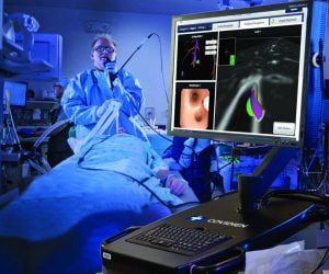
The ENB’s GPS system works like this: First, the patient’s lungs are imaged with endobrachial ultrasound, or EBUS. The crisp, highly-detailed pictures are loaded into the ENB computer, which creates a 3-D map of the patient’s lungs. Then the patient lies on a special low-frequency electromagnetic bed and is sedated for comfort.
As in a normal bronchoscopy, doctors insert a long, thin, lighted tube through the mouth and into the airways, but the ENB also has an extended wire with a locatable guide at its tip. As it travels over the electromagnetic bed, the precise whereabouts of the locatable guide inside the lungs can be seen on a bedside computer screen, much as your car’s location shows up in real time as a moving dot on your GPS or phone’s mapping app.
Using the lung and locatable guide images, doctors then slowly and carefully “drive” the ENB tip to its final destination.
In February 2014, Yant reported to Poudre Valley Hospital for his outpatient ENB procedure. Because it was the technology’s UCHealth Northern Colorado debut, a number of medical professionals observed iy. “The room was packed with people,” he recalled.
“I thought it was pretty exciting for him to be the first,” said Rose, Yant’s wife. “And it was a simple procedure. We were back home a few hours later.”
“When we combine ENB with EBUS ultrasound, we have a fantastic technology,” DePriest said. “We can accurately target a peripheral lung mass as well as the lymph nodes. A pathologist standing by in the procedure room can then tell us immediately if a mass or the lymph nodes are cancerous. In Roy’s case, only the lung tumor was malignant, so we knew he was a surgical candidate.”
Doctors of different but related specialties often work together to help UCHealth patients, DePriest said. The multidisciplinary clinic where he practices includes a thoracic surgeon, a radiologist, pulmonologists, research coordinators and a lung-patient navigator. The staff and physicians gather to discuss in detail the case of every patient on that week’s docket. They put their heads together and ask questions like: Do we have the right tissue samples? What are the best treatment options for this patient? How is the patient feeling physically? The vetted answers help ensure each patient receives the best care.
“When a person walks into something like lung cancer — that sounds like a death sentence,” DePriest said. “But when multiple experts are weighing in on each patient’s case, and we can bring the consensus recommendation to the patient, they know that an entire team is helping figure out the best approach.”
A few weeks after Yant’s ENB procedure, UCHealth thoracic surgeon Michael Stanton, MD, removed the upper lobe of Yant’s left lung. The Fort Collins resident didn’t require chemotherapy or radiation, and now he’s back to golfing, fishing and volunteering at the American Legion.
“I feel good now,” he said.
What’s ENB for?
Deployed in early 2014 at Poudre Valley Hospital, Electromagnetic Navigation Bronchoscopy, or ENB, offers patients many benefits:
• A new approach to detecting lung cancer earlier.
• Availability to a wide range of patients, including those who suffer from poor lung function or have had cancer surgery, chemotherapy, and/or radiation therapy.
• Enables physicians to take tissue samples from lung masses in areas that are inaccessible with traditional bronchoscope.
• Can be used to place fiducial markers for guiding external beam radiation therapy.
• Can be used to place markers near the lung’s surface for video-assisted thoracic surgery, or VATS.